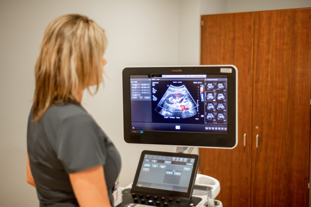
Ultrasound technology uses high-frequency sound waves and a computer to create images of blood vessels, tissues and organs of the body. Ultrasound enables the radiologist to view internal organs and examine parts of the body such as the abdomen, breasts, pelvis, thyroid and vascular system.
The exam is commonly used in conjunction with other imaging studies to confirm diagnosis or assist in the identification and biopsy of breast abnormalities. When combined with advanced elastography applications, ultrasound is particularly effective in helping to classify breast lesions and aid in the differentiation between benign and malignant tumors.
Southwoods is leading with advances in ultrasound techniques that are not available at other local imaging facilities.

© 2025 Southwoods Health. All Rights Reserved.